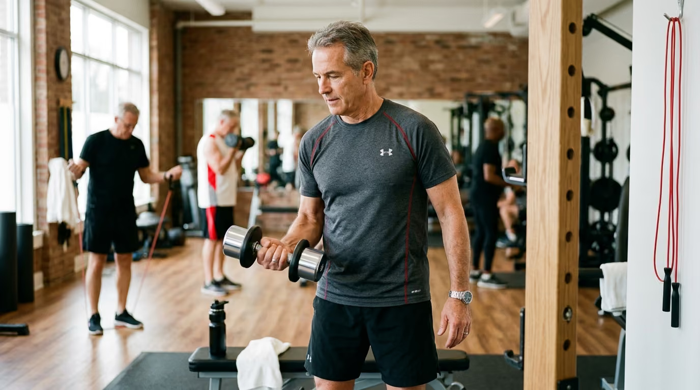
Most men believe skeletal fragility is exclusively a woman's health concern. That assumption is dangerously wrong. While women experience a rapid acceleration of bone loss during menopause, men lose bone steadily across midlife and later life. When men sustain fragility fractures, their complication and mortality rates often exceed those seen in women of the same age.
Skeletal health is not simply a matter of taking calcium pills or drinking milk. Bone is dynamic, living tissue that responds continuously to physical strain, metabolic signals, nutrition, and hormonal status. Protecting your skeleton after 45 requires understanding how mechanical loading shapes bone architecture, why secondary medical conditions cause hidden bone loss, and how to train your body to resist fractures.
Understand What The Research Shows About Bone Strength
Clinical research views bone health through the combined lenses of bone mineral density, bone quality, muscle power, and fall mechanics. Dual-energy X-ray absorptiometry, commonly known as a DXA scan, measures the amount of mineral packed into specific regions of your skeleton. This measurement generates a T-score, which compares your bone density against that of a healthy young adult. A T-score between -1.0 and -2.5 indicates osteopenia, while a score of -2.5 or lower defines osteoporosis.
Bone mineral density does not tell the entire story. Research shows that bone strength also depends on structural factors that standard DXA scans cannot see. These factors include cortical thickness, the microscopic architecture of trabecular bone, accumulated microdamage, and bone geometry. A man with mild osteopenia can sustain a fracture if his balance is poor and he takes a hard fall, while a man with lower density might avoid fractures through superior muscular control and fall resistance.
- Mechanical Load (Lifting, Impact, Ground Force)
- Osteocytes Detect Skeletal Strain
- Osteoblasts Build New Matrix Osteoclasts Resorb Old Bone
- (Mineralization & Formation) (Breakdown & Remodeling)
- Net Skeletal Strength and Density
Skeletal tissue maintains itself through a continuous cycle called remodeling. Specialized cells called osteoclasts break down old or damaged bone tissue. Another group of cells, called osteoblasts, creates a flexible protein matrix that mineralizes into fresh bone. Embedded deep inside the calcified matrix are osteocytes, which act as mechanical sensors. When you lift weights, jump, or sprint, osteocytes detect the mechanical strain and send biochemical signals that stimulate new bone formation.
Scientific models describe this adaptive response through the mechanostat principle. Much like muscular hypertrophy, bone tissue responds when physical strain exceeds a habitual threshold. When mechanical demands drop below that threshold, remodeling shifts toward resorption, and bone tissue slowly degrades. Fracture risk tools like FRAX combine bone density results with age, fracture history, parental hip fractures, smoking status, and medication history to estimate ten-year fracture probabilities.
Examine Why Skeletal Remodeling Changes After 45
After age 45, the balance between bone breakdown and bone rebuilding shifts. Osteoblasts become less efficient at replacing the matrix removed by osteoclasts. Over time, each remodeling cycle leaves a small deficit. Cortical bone becomes thinner and more porous, while the internal trabecular meshwork loses connectivity.
Declining physical activity accelerates this biological shift. When daily movement decreases, the mechanical signals that trigger osteocytes weaken. Sarcopenia, the age-related loss of muscle mass and contractile strength, reduces the internal forces applied to bones during movement. Because muscle contractions exert the largest physiological loads on the skeleton, falling muscle power directly lowers the stimulus for bone maintenance.
- Aging Pathways After 45
- • Reduced Mechanical Load Weaker osteocyte strain signaling
- • Loss of Muscle Mass Lower internal tension on bone attachments
- • Hormonal Shifts Lower androgen conversion to estradiol
- • Secondary Factors Medication side effects, alcohol, malabsorption
Hormonal alterations play a distinct role in male skeletal health. In men, testosterone maintains bone mass partly through direct androgen receptor activation, but largely through aromatization into estradiol. Circulating estrogen is critical for regulating bone resorption in men. As total and bioavailable testosterone concentrations decline with age, local estradiol synthesis within bone tissue also decreases, allowing osteoclasts to resorb bone at a faster rate.
Secondary medical causes account for up to 60 percent of male osteoporosis cases. Prolonged exposure to glucocorticoid medications, such as prednisone, directly suppresses osteoblast function and reduces calcium absorption. Androgen deprivation therapy for prostate cancer eliminates circulating androgens and triggers rapid, severe bone demineralization. Other common secondary drivers include heavy alcohol consumption, cigarette smoking, chronic kidney disease, hyperthyroidism, hyperparathyroidism, and malabsorption disorders like celiac disease.
For a deeper look into the systemic changes that occur during midlife, read our resource on longevity science and health optimization.
Translate Clinical Data Into Daily Capability and Function
A loss of bone mass produces no pain or obvious outward symptoms until a fracture occurs. This silent progression makes bone health easy to ignore until a routine slip on an icy driveway or a stumble off a curb leads to a broken hip or wrist. Fragility fractures result from low-energy trauma, defined as forces equal to or less than a fall from standing height.
Sustaining a fragility fracture alters physical capability, mobility, and independence. Hip fractures in older men require major surgery and extensive rehabilitation, and research indicates that up to a third of men die within twelve months following a hip fracture. Many survivors never regain their previous walking speed, balance, or ability to live independently without assistance.
- Daily Capability Link
- Lower Bone Quality Loss of Balance Fall Event Fragility Fracture
- High Muscle Power Robust Skeletal Load Fall Resistance Maintained Independence
Vertebral fractures are the most common osteoporotic fractures in men, yet they frequently go undiagnosed. These spinal compression fractures often happen during ordinary activities like lifting a heavy box, shoveling snow, or bending forward abruptly. They can present as dull, nagging back pain rather than sharp agony. As multiple vertebral bodies collapse, men experience height loss, a rounded upper back known as kyphosis, restricted lung capacity, and chronic muscular fatigue.
Preserving physical function requires treating bone density, muscle power, and balance as a single unified system. You need skeletal stiffness to support heavy loads, muscular strength to move your joints, and neuromuscular coordination to prevent falls. Maintaining these attributes lets you hike, lift, travel, and play sports late in life without fear of sudden skeletal breakdown.
If you are working to maintain overall functional performance as you age, explore our dedicated guides on healthy aging strategies.
Apply Progressive Mechanical Loading and Resistance Training
Bone tissue requires specific, dynamic loading to stimulate remodeling. Research consistently demonstrates that passive weight-bearing or steady-state walking is insufficient on its own to build bone density in the spine and hips. Walking provides valuable cardiovascular benefits, but its loading rates are too low and predictable to prompt significant osteogenic adaptation.
To stimulate bone remodeling, an exercise must satisfy four loading rules:
- It must produce high strain magnitude by challenging muscles against heavy resistance.
- It must deliver a high strain rate through rapid force development or impact.
- It must apply forces in diverse, non-habitual directions.
- It must progress over time as the skeleton adapts.
- Loading Principle Breakdown
- • Heavy Resistance High muscular tension pulls on tendon-bone junctions
- • Impact Forces Ground reaction waves travel through hip and spine
- • Novel Directions Multi-planar movements challenge weak skeletal zones
- • Progressive Load Increasing resistance prevents bone adaptation plateaus
Progressive resistance training forms the foundation of skeletal preservation. Exercises that load the axial skeleton and hips yield the greatest improvements in bone mineral density. Multi-joint compound lifts, including squats, deadlifts, overhead presses, and loaded carries, apply heavy compressive and tensile forces across the spine, femoral neck, and pelvis. Systematic reviews indicate that both heavy-load training and supervised moderate-load training can elicit positive bone responses, provided sets are taken close to muscular fatigue.
Impact training introduces rapid loading rates that send shockwaves through the skeleton. Activities like jumping rope, box jumps, broad jumps, and brisk multidirectional footwork stimulate cortical bone thickening. For men with healthy joints and no prior fractures, adding brief sets of high-impact jumps to a warm-up provides a strong osteogenic stimulus. Sedentary men or those returning to exercise should introduce impact training gradually to allow connective tissues and bone matrix time to adapt.
To structure a safe lifting routine that supports bone and joint health, review our core principles on strength and muscle development.
Optimize Nutrition, Mineral Intake, and Hormonal Health
Building and maintaining skeletal tissue requires a continuous supply of structural minerals and dietary protein. Calcium forms the primary mineral crystal, hydroxyapatite, which gives bone its compressive rigidity. Clinical guidelines from the Endocrine Society and the Bone Health and Osteoporosis Foundation recommend that men consume between 1,000 and 1,200 milligrams of calcium daily.
Dietary sources are the preferred way to meet your calcium requirements. Dairy products like yogurt, milk, and hard cheeses supply bioavailable calcium alongside protein. Non-dairy sources include calcium-set tofu, canned sardines or salmon with edible bones, and fortified plant milks. When your daily diet falls short, low-dose calcium supplements can bridge the gap. Avoid taking massive single doses of supplemental calcium, because excessive supplementation offers no additional bone benefit and has been linked to potential vascular complications.
- Nutritional Support Checklist
- • Total Calcium: 1,000 to 1,200 mg daily (prioritize dairy, canned fish, fortified foods)
- • Vitamin D: Maintain serum 25(OH)D levels at or above 30 ng/mL
- • Dietary Protein: 1.2 to 1.6 grams per kilogram of body weight
- • Whole Foods: Provide magnesium, vitamin K2, and essential micronutrients
Vitamin D is necessary for intestinal calcium absorption and bone mineralization. Without adequate vitamin D, your body cannot absorb dietary calcium efficiently, forcing parathyroid hormone levels to rise and strip calcium from your bones. Clinical guidelines suggest maintaining blood levels of 25-hydroxyvitamin D at or above 30 ng/mL. Many men over 45 require daily supplementation of 800 to 2,000 IU, depending on sun exposure, skin tone, geographical location, and baseline blood values.
Bone is roughly fifty percent protein by volume. The internal collagen scaffolding provides tensile strength, allowing bone to bend slightly under load without shattering. Consuming 1.2 to 1.6 grams of protein per kilogram of body weight daily supports bone matrix renewal while preserving lean muscle mass. Severe calorie restriction and crash dieting accelerate bone loss by reducing IGF-1 signaling, lowering sex hormones, and degrading bone collagen.
For an extensive review of hormonal balance and endocrine vitality, see our resources on hormones and male vitality.
Modify Movement Patterns and Avoid High-Risk Spinal Strains
A diagnosis of osteopenia or osteoporosis does not mean you should stop lifting weights or avoiding active movement. Complete rest and inactivity accelerate bone demineralization and muscle wasting. The solution is to modify high-risk movement patterns while continuing to load your skeleton safely.
The primary movement risk for individuals with low bone density is loaded, rapid, or end-range spinal flexion. Bending forward at the waist while rounding your lower back places extreme compressive force on the anterior edges of your vertebral bodies. When combined with rotational twisting, this position dramatically increases the risk of a spinal compression fracture.
- Movement Modification Guide
- • High-Risk Pattern: Loaded, rapid spinal flexion (e.g. standard sit-ups, toe touches)
- Safe Replacement: Hip-hinge mechanics, neutral-spine romanian deadlifts
- • High-Risk Pattern: End-range twisting under external load (e.g. seated torso twists)
- Safe Replacement: Anti-rotation holds, Pallof presses, suitcase carries
- • High-Risk Pattern: Uncontrolled explosive jumping with existing fractures
- Safe Replacement: Heavy sled pushes, controlled step-ups, brisk incline walks
Learn to separate hip movement from spinal movement. Master the hip hinge, keeping your spine neutral while your hips travel backward during lifting and daily bending. Replace traditional sit-ups and toe-touch stretches with core stability exercises like planks, bird-dogs, and Pallof presses that resist unwanted trunk motion.
If you have a history of vertebral compression fractures or severe osteoporosis, avoid high-impact jumping and aggressive spinal rotation. Substitute these with controlled resistance movements, neutral-spine cable rows, step-ups, and brisk walking. Working with a physical therapist familiar with osteoporosis guidelines will help you build a personalized exercise program that protects vulnerable areas.
For practical advice on joint protection and safe movement mechanics, review our guides on mobility and recovery.
Screen Early and Navigate Clinical Conversations
Osteoporosis screening in men remains inconsistent across standard medical practice. The Bone Health and Osteoporosis Foundation and the Endocrine Society recommend routine DXA screening for all men aged 70 and older. They also recommend screening men aged 50 to 69 who display clinical risk factors. In contrast, the United States Preventive Services Task Force concluded that current evidence is insufficient to assess the balance of benefits and harms of universal screening in men.
Because universal screening is not standard for men in their fifties and sixties, you must be your own advocate. Discuss a DXA scan with your physician if you have any key risk factors. These include a prior low-trauma fracture, long-term steroid use, androgen deprivation therapy, low body weight, heavy alcohol use, or a family history of hip fractures.
- Clinical Evaluation Steps
- 1. Schedule a baseline DXA scan (hip, femoral neck, and lumbar spine).
- 2. Calculate your FRAX score to establish ten-year absolute fracture risk.
- 3. Review prescription medications for bone-depleting side effects.
- 4. Screen for secondary conditions
- • Total and free testosterone, LH, and prolactin
- • Serum calcium, phosphate, and parathyroid hormone (PTH)
- • 25-hydroxyvitamin D
- • Thyroid stimulating hormone (TSH)
- • 24-hour urinary calcium excretion
- • Complete blood count and comprehensive metabolic panel
- 5. Request lateral thoracic and lumbar imaging if height loss or back pain is present.
When discussing bone health with your physician, ask clear questions about secondary testing. If your DXA scan reveals osteopenia or osteoporosis, do not settle for a basic calcium recommendation. Ask your doctor to investigate underlying causes, including hypogonadism, malabsorption, kidney disease, or parathyroid dysfunction. Identifying and treating a secondary condition often halts bone loss without requiring immediate osteoporosis medications.
Always consult a qualified healthcare provider regarding medical testing and personalized treatment decisions. You can read our complete health and medical disclaimer for additional context on our editorial standards.
Discard Common Misreadings About Male Skeletal Health
Several persistent myths prevent men from taking effective action to protect their skeletons. Relying on outdated advice increases your risk of avoidable fractures.
The first misconception is that osteoporosis is purely a women's health problem. While women develop the condition in greater numbers, one in four men over age 50 will experience an osteoporotic fracture in their lifetime. Men are also far less likely to be screened or prescribed protective therapies after breaking a bone, leading to worse long-term outcomes.
The second misconception is that regular walking is all you need to keep your bones strong. Walking offers substantial cardiovascular and metabolic benefits, but it does not generate enough mechanical force to stimulate bone formation in your spine or hips. You must combine walking with progressive resistance training and multi-directional loading to stimulate bone remodeling.
- Common Myth vs. Clinical Reality
- • Myth: "Taking high-dose calcium pills guarantees strong bones."
- Reality: Calcium supplies raw mineral, but mechanical strain directs bone formation.
- • Myth: "Diagnosed bone loss means you should never lift weights again."
- Reality: Progressive, neutral-spine lifting strengthens bone and reduces fall risk.
- • Myth: "Low testosterone always requires immediate hormone replacement for bone health."
- Reality: True hypogonadism must be confirmed, and other bone-specific treatments evaluated.
The third misconception is that massive calcium supplementation is harmless insurance. Your intestines can only absorb a limited amount of calcium at one time, and excess supplemental calcium does not increase bone density. Focus on hitting 1,000 to 1,200 milligrams daily through food first, using small supplemental doses only when your diet falls short.
The fourth misconception is that a diagnosis of osteopenia means you must avoid the weight room. Avoiding resistance training leads to rapid muscle loss, poorer balance, and accelerated bone thinning. By learning proper lifting form, avoiding loaded spinal flexion, and progressing gradually, resistance training remains one of your most effective tools for skeletal defense.
Recognize Where Scientific Evidence Remains Incomplete
While the core principles of bone remodeling and mechanical loading are well established, clinical research still contains gaps regarding male osteoporosis. Knowing where the evidence is limited helps you avoid unproven treatments and commercial hype.
First, optimal exercise prescription parameters for men with established osteoporosis remain incompletely defined. Most large-scale exercise trials have been conducted in postmenopausal women. While researchers know that heavy resistance and impact stimulate bone, the exact combination of sets, reps, and loading frequencies that maximizes bone growth in older men without triggering joint flare-ups requires further investigation.
Second, the clinical utility of routine testosterone replacement therapy solely for fracture prevention remains unclear. While testosterone therapy increases bone mineral density in men with documented, symptomatic hypogonadism, clinical trials have not proven that testosterone therapy alone reduces long-term fracture rates in men with normal or borderline hormone levels.
- Evidence Grading in Male Bone Research
- • Resistance Training & Impact: Strong evidence for improving BMD and physical function.
- • Calcium Vitamin D Synergy: Strong evidence for maintaining mineral supply.
- • Universal DXA Screening in Men Under 70: Mixed recommendations across major guidelines.
- • Standalone Megadose Vitamin D: Weak evidence for fracture reduction.
- • Bone Density Drugs in Men: Strong evidence for specific bisphosphonates and anabolics.
Third, the effect of non-weight-bearing endurance sports on male bone density requires careful interpretation. Observational studies show that dedicated road cyclists and swimmers often have lower bone mineral density than runners or strength athletes, even when their cardiovascular fitness is elite. However, randomized trials exploring the best supplemental training routines to reverse bone loss in competitive master cyclists are still ongoing.
Finally, evidence for standalone high-dose vitamin D supplementation in the absence of deficiency is weak. Clinical trials have found that giving massive, infrequent boluses of vitamin D does not reduce fracture risk and can even increase fall rates in older adults. Vitamin D works best as part of a steady, daily nutritional strategy combined with adequate calcium and physical training.
Key Takeaways
- Bone health depends on a combination of bone density, bone quality, muscle power, balance, and fall prevention.
- Secondary causes, such as steroid medications, hypogonadism, alcohol use, and gastrointestinal malabsorption, drive up to 60 percent of male osteoporosis cases.
- Steady-state walking alone does not provide sufficient mechanical force to build or maintain bone mineral density in your hips and spine.
- Progressive resistance training and safe impact exercise supply the primary mechanical signals needed to stimulate bone remodeling.
- Target 1,000 to 1,200 milligrams of daily calcium primarily from food, and maintain blood 25-hydroxyvitamin D levels at or above 30 ng/mL.
- Protect your spine by maintaining a neutral posture during heavy lifts and avoiding loaded, rapid spinal flexion or extreme twisting under load.
- Men over 70, and men over 50 with clinical risk factors, should actively request a DXA scan and FRAX risk calculation from their doctor.
Protecting your skeleton after 45 requires combining progressive resistance training, targeted bone nutrition, and proactive clinical screening to keep your body strong, durable, and fully capable for life.
Sources
- Osteoporosis in Men: An Endocrine Society Clinical Practice ...
- Strong, steady and straight: UK consensus statement on physical activity and exercise for osteoporosis
- Exercise for individuals with osteoporosis
- Exercise for preventing bone loss and reducing fracture risk
- The Central Role of Osteocytes in the Four Adaptive Pathways of Bone's Mechanostat - PubMed
- Exercise: Preventing bone loss and reducing fracture risk
- Strong, steady and straight: UK consensus statement on physical activity and exercise for osteoporosis
- Exercise and physical activity in individuals at risk of fracture
- Mechanical regulation of bone remodeling | Bone Research
- 1
- Exercising with osteoporosis: Stay active the safe way
- Male osteoporosis: pathophysiology, risk assessment, and ...
- Osteoporosis in Males - StatPearls - NCBI Bookshelf
Stay sharp
Get the word
Join our newsletter for the best writing advice and stories from the field.