Joint longevity is not the complete absence of wear, nor is it the pursuit of a flawless magnetic resonance scan. It is the preservation of usable physical capacity across decades. True joint capacity means your knees, hips, shoulders, and spine can handle the forces of daily life, recreational sport, and physical training without persistent pain or loss of independence.
It is not built through passive treatments, resting indefinitely when aches appear, or relying on unverified joint supplements. It requires a balanced combination of progressive strength, regular movement across varied angles, sensible load management, and an understanding of normal aging.
This guide examines the scientific evidence behind joint capacity for men over 45. It outlines the mechanics of the major joints, breaks down how body weight and recovery interact with physical loading, and details how to distinguish standard mechanical sensations from issues that require medical evaluation.
What Does the Research Show About Maintaining Joint Capacity?
The strongest scientific evidence on joint health points to regular physical activity and progressive resistance training. Major health organizations do not recommend bed rest or joint immobilization for maintaining physical function. The World Health Organization recommends that all adults complete 150 to 300 minutes of moderate aerobic activity weekly, or 75 to 150 minutes of vigorous activity.
The World Health Organization also advises performing muscle strengthening exercises for all major muscle groups at least two days per week. For older adults, the guidance adds multicomponent training focusing on functional balance and strength three or more days per week. This combination preserves the ability to walk, stand up from low chairs, navigate uneven terrain, and recover from unexpected trips.
Clinical practice guidelines from rheumatology organizations support exercise as a foundational intervention for joint conditions. The American College of Rheumatology and the Arthritis Foundation recommend exercise and physical activity for hip and knee osteoarthritis. Research reviews analyzing physical therapy outcomes consistently show that local muscular strength and aerobic fitness reduce pain and improve physical function.
Strength serves as a capacity reserve for your joints. When your quadriceps, hamstrings, and gluteal muscles are strong, they absorb and disperse ground reaction forces during walking or stair climbing. That strength reduces the mechanical burden placed directly on cartilage and subchondral bone.
The evidence does not show that physical activity or recreational sports cause joint degeneration in healthy individuals. A systematic review investigating midlife physical activity found no consistent link between cumulative exercise and the development of knee osteoarthritis. Previous traumatic joint injury, high occupational loads, and obesity were the primary risk factors identified by researchers.
Running offers a clear example of how physical loading affects joints. A large systematic review and meta-analysis analyzed the prevalence of hip and knee osteoarthritis across different activity levels. The researchers found an osteoarthritis rate of 3.5 percent in recreational runners, compared to 10.2 percent in sedentary controls. Competitive, elite runners who sustained exceptionally high mileage over decades showed a higher prevalence of 13.3 percent.
This data indicates that moderate, recreational loading is associated with healthy joint tissues rather than accelerated deterioration. Joints adapt to applied loads when those loads are introduced systematically and matched with adequate recovery.
- JOINT CAPACITY MANAGEMENT SPECTRUM
- UNDERLOADING OPTIMAL ZONE OVERLOADING
- Tissue deconditioning - Progressive load - Sudden spikes
- Reduced tolerance - Adequate recovery - Poor recovery
- Increased sensitivity - Capacity reserve - Chronic pain
Sensible load management requires keeping regular exposure while avoiding sudden spikes in volume or intensity. Complete inactivity causes tendons, muscles, and joint structures to decondition rapidly. When deconditioned tissues are suddenly exposed to high demands, the risk of irritation rises.
Gradual progression allows tissues to remodel. A reliable training plan increases variables such as weight, repetitions, speed, or movement range in small increments rather than large leaps.
Range of motion is valuable when it supports practical tasks. A capable hip requires enough flexion and rotation to squat, sit comfortably, and walk without compensation. A functional shoulder requires elevation and rotation to reach overhead and carry loads without neck strain.
Current research does not establish a universal range of motion number that guarantees protection against joint stiffness. The priority is developing comfortable, controlled movement through ranges required for your personal activities and training goals.
Why Do Joints Change After 45?
Joint changes after age 45 reflect normal biological shifts rather than a disease process. Understanding these changes helps men make intelligent adjustments to their training without fear or frustration.
Cartilage undergoes subtle composition changes over time. Articular cartilage is composed of water, collagen, and proteoglycan molecules. With age, the water content within the extracellular matrix shifts, and the rate of proteoglycan synthesis decreases.
These changes reduce the fluid pressure mechanism that helps cartilage disperse compressive forces. The tissue remains durable, but it responds better to gradual warm-ups and progressive loads than to sudden, cold impacts.
Tendons and ligaments also experience alterations in collagen turnover. Tendon stiffness and cross-linking change with age, which can reduce the elastic energy storage capacity of the tissue. Tendons require slightly longer to adapt to changes in training volume or high-velocity movements.
Muscular changes occur alongside these connective tissue shifts. Sarcopenia, the age-related reduction in skeletal muscle mass and muscle fiber diameter, begins gradually in midlife.
Type II fast-twitch muscle fibers experience a faster rate of decline than Type I endurance fibers. Fast-twitch fibers provide rapid force production to stabilize a knee during a slip or catch a heavy load. Maintaining these fibers through strength and muscle training principles protects the surrounding joint structures from unexpected stress.
Synovial fluid production responds directly to joint movement. Synovial fluid lubricates the articular surfaces and delivers nutrients to cartilage, which lacks a direct blood supply. When daily movement becomes restricted to sedentary postures, the distribution of synovial fluid decreases.
Regular movement cycles synovial fluid through the joint space. This fluid exchange reduces stiffness and supports cartilage metabolism throughout midlife and older age.
Hormonal and metabolic factors also influence joint tissues after 45. Gradual shifts in systemic hormone levels affect protein synthesis and tissue remodeling rates. Low-grade systemic inflammation, often linked with metabolic changes, can increase pain sensitivity in peripheral joints.
Joint changes after 45 are not a sign of physical fragility. They simply mean that tissue recovery and adaptation require consistent physical signals, good sleep, and structured training progression.
What Does Joint Capacity Mean in Daily Life and Training?
In daily life, joint capacity is the difference between your maximum physical capability and the demands of your routine activities. When your physical capacity is high, ordinary tasks feel effortless and carry a low risk of strain.
Consider the act of walking down a steep flight of stairs or descending a hiking trail. Descending places an eccentric load on the quadriceps and patellar tendon equal to several times your body weight. A man with strong leg muscles and stable knees absorbs that force smoothly through muscular contraction.
A man with low muscular reserve must rely more heavily on passive joint structures to absorb the downward force. Over a long descent, this reliance can lead to anterior knee irritation and joint stiffness.
Carrying heavy objects provides another practical example of joint capacity. Lifting a 40-pound bag of soil or loading luggage into an overhead compartment requires coordination between the spine, hips, and shoulders.
The hips must hinge to distribute force through the glutes and hamstrings. The spine must transfer force through the core musculature. The shoulder requires stable scapular movement to position the arm without pinching soft tissue.
When joint capacity declines, men often modify their lives unconsciously. They begin avoiding stairs, skipping hikes with family, or abandoning favorite sports because their joints feel unpredictable.
This avoidance creates a downward cycle. Less activity leads to weaker muscles, stiffer tendons, and further reductions in joint tolerance.
Maintaining capacity breaks this cycle. When you build strength and preserve movement quality, you maintain the freedom to travel, participate in sports, and handle manual tasks with confidence.
Joint capacity also provides a safety margin for unexpected physical accidents. Stepping into an unseen hole in the yard or slipping on wet pavement requires immediate joint stabilization.
Strong hips, reactive calves, and a responsive trunk can correct your balance before a fall occurs. If a minor twist does occur, conditioned tissues recover faster than deconditioned tissues.
Viewing joint health through the lens of usable capacity shifts your focus from avoiding movement to building physical capability.
How Should You Manage Movement for Knees, Hips, Shoulders, and Spine?
Each major joint complex has specific structural roles and mechanical needs. Balancing stability and mobility across these four areas maintains full-body function.
- JOINT ROLES AND TRAINING TARGETS
- JOINT REGION PRIMARY ROLE KEY MUSCLE GROUPS
- Knee Stable hinge Quadriceps, hamstrings, calf
- Hip Multi-planar mobile Gluteals, adductors, rotators
- Shoulder Dynamic mobility Rotator cuff, scapular beds
- Spine Segmental transfer Core trunk, erectors, lats
The Knee: Preserving a Powerful Hinge
The knee functions primarily as a hinge joint designed to handle large compressive and shear forces during locomotion. It relies heavily on the hip above and the ankle below for proper alignment.
When hip rotators or ankle joints lack mobility, the knee often experiences excess rotational torque. Training the knee requires building strength throughout the quadriceps, hamstrings, and calves while maintaining stable tracking.
Key strategies for knee capacity include:
- Progressive squatting and stepping: Squats, split squats, and step-ups strengthen the quadriceps and improve patellofemoral load tolerance.
- Hamstring conditioning: Romanian deadlifts and leg curls build posterior strength, protecting the anterior cruciate ligament.
- Calf and soleus loading: Standing and seated calf raises strengthen the lower leg muscles that absorb impact during walking and running.
- Gradual range exposure: Moving through deeper knee flexion under light loads preserves cartilage tolerance at full joint angles.
The Hip: Developing Multi-Directional Strength
The hip is a robust ball-and-socket joint built for both power and mobility. It requires strength across multiple planes: flexion, extension, abduction, adduction, and rotation.
Sedentary desk work often leaves the hip flexors short and the gluteal muscles underutilized. Restoring hip capacity reduces stress on both the lumbar spine and the knee joints.
Key strategies for hip capacity include:
- Hip hinging movements: Deadlifts, kettlebell swings, and hip thrusts build powerful gluteal and hamstring force production.
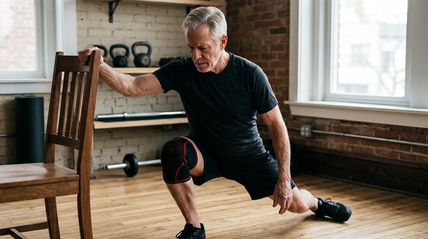
- Lateral and rotational work: Lateral lunges, banded side steps, and rotational drills strengthen the gluteus medius and deep hip rotators.
- End-range extension: Lunges and split stances stretch the anterior hip while engaging the rear glute.
- Controlled mobility: Seated or standing hip rotations preserve the internal and external rotation needed for comfortable walking.
The Shoulder: Balancing Mobility and Stability
The shoulder is the most mobile joint complex in the human body, relying on the shallow glenohumeral socket and the moving scapula. Shoulder longevity depends on the coordination between the large prime movers and the stabilizing rotator cuff muscles.
Pressing heavy weights without adequate pulling volume can disrupt this balance, leading to impingement sensations or tendon irritation. Exploring structured mobility and recovery resources can help balance upper body joint demands.
Key strategies for shoulder capacity include:
- Balanced pulling and pushing: Pair overhead and horizontal pressing with equal or greater volumes of rowing and face pulls.
- Scapular control: Movements like serratus wall slides and scapular pull-ups ensure the shoulder blade rotates upward smoothly during reaching.
- Rotator cuff strengthening: External and internal rotations with bands or light dumbbells build muscular endurance in stabilizing tendons.
- Loaded carries: Farmer walks and suitcase carries challenge the shoulder girdle to maintain stability under continuous load.
The Spine: Supporting Force Transfer and Controlled Motion
The spine is a segmented column designed to protect the central nervous system while allowing flexion, extension, side-bending, and rotation. Long-term spinal health does not require keeping the back completely rigid at all times.
Instead, the spine needs the capacity to brace against heavy external loads and move fluidly through daily tasks. Building strength in the spinal erectors, abdominal wall, and latissimus dorsi provides full-torso support.
Key strategies for spinal capacity include:
- Core bracing patterns: Planks, bird-dogs, and Pallof presses train the trunk to resist unwanted movement under load.
- Controlled spinal articulation: Unloaded movements like cat-cow drills maintain comfortable segmental motion throughout the vertebrae.
- Hip-dominant lifting: Learning to hinge at the hips during heavy lifting spares the lumbar spine from excessive shearing forces.
- Rotational strength: Woodchops and cable rotations train the oblique musculature to control and produce rotational power safely.
What Role Do Body Weight and Recovery Play in Long-Term Joint Health?
Mechanical loading and systemic biology interact continuously to influence joint comfort. Body weight and recovery practices represent two major factors in this relationship.
Body mass affects the compressive forces transmitted through weight-bearing joints like the knees and hips. During level walking, the force passing through the knee joint is roughly two to three times your body weight.
When climbing stairs or jogging, that force can rise to four to six times your body weight. An increase in body weight naturally magnifies these cumulative mechanical forces across millions of steps per year.
Scientific research demonstrates a clear link between elevated body mass index and knee osteoarthritis risk. A systematic review and meta-analysis indexed in PubMed calculated a pooled odds ratio of 2.63 for knee osteoarthritis in individuals with obesity compared to normal-weight peers.
A large cohort analysis found that every 5-unit increase in body mass index was associated with a 35 percent rise in knee osteoarthritis risk.
Body weight is not purely a mechanical issue. Adipose tissue functions as an active endocrine organ that releases pro-inflammatory signaling proteins called adipokines.
These circulating compounds can contribute to low-grade systemic inflammation throughout the body. This systemic effect helps explain why elevated body mass is also associated with joint discomfort in non-weight-bearing joints, such as the hands.
For individuals with existing joint symptoms and elevated body mass, modest weight loss provides measurable relief. Clinical reviews show that losing 5 to 10 percent of total body weight produces significant improvements in joint pain, self-reported disability, and physical function.
Losing more than 10 percent of body weight yields even larger gains in physical capacity. A prospective cohort study observed that adults who reduced initial body weight by over 7.5 percent had lower rates of subsequent total knee replacement.
- EFFECTS OF WEIGHT REDUCTION ON JOINTS
- 5% to 10% WEIGHT REDUCTION 10% WEIGHT REDUCTION
- Measurable pain reduction - Substantial pain relief
- Improved daily function - Major functional gains
- Reduced systemic markers - Lower joint stress loads
Weight management should never compromise muscle mass. Aggressive dieting without adequate protein and resistance exercise causes the loss of valuable skeletal muscle.
Losing muscle reduces the body's active shock absorbers, leaving joints more vulnerable to mechanical fatigue. The priority must always be preserving lean muscle while reducing excess body fat.
Recovery practices allow joint tissues to adapt to physical demands. Cartilage, tendons, and ligaments have lower metabolic rates and blood flow than skeletal muscle, meaning they adapt on a slightly longer timeline.
While science has not established a precise number of recovery hours required for joint longevity, physical indicators provide clear guidance. A well-recovered joint system displays predictable patterns:
- Muscular soreness resolves within 24 to 48 hours following a training session.
- Joint stiffness in the morning lasts less than 15 to 30 minutes.
- Movement range remains consistent from week to week without progressive tightening.
- Physical strength and movement control remain steady or improve over time.
- No accumulation of localized swelling, warmth, or persistent resting pain occurs.
When recovery is insufficient, joints become irritable. If training sessions consistently produce escalating pain or next-day movement restrictions, the overall load exceeds current tissue tolerance. Adjusting volume, exercise selection, or rest intervals allows tissues to remodel successfully.
What Practical Training Habits Build Joint Capacity After 45?
Building durable joints after 45 requires structured training habits rather than random workouts. Applying established training principles ensures consistent progress while minimizing irritation.
- JOINT LONGEVITY TRAINING HABITS
- 1. Dynamic Preparation 5 to 10 minutes of active movement
- 2. Progressive Resistance 2 to 3 weekly multi-joint sessions
- 3. Movement Variability Varied planes, angles, and tempos
- 4. Auto-Regulation Modify loads based on joint feedback
- 5. Functional Balance Single-leg work and stability drills
1. Prioritize Dynamic Preparation
Cold, sedentary joints do not respond well to immediate heavy loads. Spend 5 to 10 minutes performing dynamic preparation before lifting or intense aerobic work.
Incorporate bodyweight squats, lunges with rotation, arm circles, and band pull-aparts. This active preparation raises tissue temperature, increases blood flow to stabilizing muscles, and coats cartilage surfaces with synovial fluid.
2. Emphasize Multi-Joint Compound Movements
Multi-joint exercises train the body to distribute mechanical forces across multiple muscle groups and joints simultaneously. Movements such as trap-bar deadlifts, goblet squats, overhead dumbbell presses, and cable rows build coordinated full-body capacity.
Perform these compound exercises through a comfortable, pain-free range of motion. Gradually increase the load over weeks and months to stimulate bone density and connective tissue remodeling.
3. Incorporate Movement Variability
Repeating the exact same exercise, stance, and load path indefinitely can create localized tissue stress. Introduce subtle variations into your weekly routine to expose joints to different angles.
Switch between barbell lifts, dumbbell variations, cable machines, and bodyweight exercises. Vary your foot stance on leg presses or alter your grip width during pull-downs.
This exposure builds broad structural capacity rather than narrow, pattern-specific tolerance. You can find detailed routines in our mobility and joint function guide.
4. Practice Auto-Regulation
Auto-regulation is the practice of adjusting your training intensity based on real-time feedback. Some days your joints will feel capable of handling heavier weights and higher volumes.
On other days, lingering fatigue, poor sleep, or minor joint stiffness will signal a need for caution. When a specific movement feels irritable, reduce the load, shorten the range of motion, slow the tempo, or swap the exercise for a joint-friendly alternative.
5. Train Balance and Single-Leg Stability
Joint capacity requires dynamic stability when supporting your body on one leg. Single-leg exercises challenge the stabilizing muscles around the foot, ankle, knee, and hip to work together.
Include single-leg Romanian deadlifts, step-ups, split squats, and single-arm carries in your weekly program. These movements improve proprioception, the body's ability to sense its position in space, which helps prevent falls and awkward joint twists.
Which Common Beliefs About Joint Health Are Not Supported by Science?
Several widespread assumptions regarding joint health and aging lack scientific backing. Examining these misconceptions prevents unnecessary fear and activity restriction.
Misconception 1: Running Automatically Destroys Knee Cartilage
A common belief is that the repetitive impact of running inevitably wears away knee cartilage and leads to joint replacement. Long-term cohort studies and systematic reviews do not support this claim for recreational runners.
Recreational runners show lower rates of knee osteoarthritis than sedentary individuals. Cartilage is a living tissue that responds to cyclic, dynamic loading by adapting its metabolic activity.
Excessive running combined with structural deformities or previous severe injuries can increase risk. However, sensible recreational running within a person's recovery capacity is safe for healthy joints.
Misconception 2: Joint Pain Means You Should Stop Moving Completely
Many men believe that experiencing joint discomfort is a signal to stop all physical exercise until the ache disappears entirely. Complete rest leads to rapid muscle atrophy, reduced synovial fluid circulation, and decreased tendon capacity.
Deconditioned joints become more sensitive to normal daily loads when activity resumes. Clinical practice guidelines for joint health strongly advocate for continued low-impact movement and modified strength training.
The goal is to modify the movement to a tolerable level, not to eliminate physical loading entirely.
Misconception 3: Magnetic Resonance Scans Tell the Whole Story
Another common assumption is that structural findings on an X-ray or scan dictate how a joint feels and functions. Medical imaging frequently shows age-related changes, such as disc bulges, cartilage thinning, or meniscus tears, in people who have zero pain or physical limitations.
Conversely, individuals with significant joint discomfort may show only minor changes on imaging. The National Institute for Health and Care Excellence advises against using routine imaging to guide standard joint care.
Treatment and physical training should be guided by your actual symptoms, strength, and functional capability rather than imaging findings alone.
Where Is the Joint Longevity Evidence Still Thin or Mixed?
While the benefits of general exercise and strength training are well-established, certain popular joint preservation concepts have limited scientific backing. Recognizing where evidence remains thin prevents wasting time and resources on unproven methods.
- EVIDENCE STRENGTH ON JOINT INTERVENTIONS
- SOLID EVIDENCE THIN OR MIXED EVIDENCE
- Progressive strength training - Joint health supplements
- Regular aerobic activity - Universal load formulas
- 5-10% weight loss for OA - Precise recovery scores
- Functional balance exercises - Extreme flexibility routines
Dietary joint supplements represent an area where evidence is mixed or weak. Products such as glucosamine, chondroitin, collagen peptides, and various botanical extracts are widely marketed for joint cartilage restoration.
Large-scale systematic reviews show inconsistent clinical benefits for these supplements, with effects often matching placebos. While these compounds are generally safe, high-quality evidence does not support them as primary tools for joint longevity.
Specific weekly load-progression percentages also lack universal scientific support. Popular training rules, such as never increasing weekly mileage or training volume by more than 10 percent, are general heuristics rather than validated laws.
Tissue tolerance depends on sleep, nutrition, training history, and life stress. Rigid mathematical rules cannot replace attentive auto-regulation and symptom monitoring.
The concept of precise recovery metrics for joint longevity is another area lacking deep evidence. Wearable devices claim to measure joint recovery through heart rate variability and sleep tracking algorithms.
These devices provide general information about autonomic nervous system balance, but they cannot assess local joint tissue status or cartilage remodeling rates. Observable functional capability remains the most reliable measure of joint readiness.
Universal flexibility targets also lack strong research support for preventing joint degeneration. While maintaining functional movement is valuable, stretching beyond the requirements of your daily activities and sports does not confer extra joint protection. Controlled strength throughout your usable range is far more protective than passive, extreme flexibility.
For deeper investigations into research-backed longevity methods, see our overview of longevity science and optimization research.
What Are the Key Warning Signs That Warrant Medical Assessment?
Most mechanical joint aches improve with sensible load adjustments, strength training, and temporary modifications. Certain symptoms, however, indicate underlying conditions that require prompt professional evaluation.
The National Institute for Health and Care Excellence outlines a standard clinical presentation for common osteoarthritis: age 45 or older, activity-related joint discomfort, and morning stiffness lasting under 30 minutes.
Symptoms that diverge from this pattern warrant clinical assessment to rule out inflammatory arthritis, infection, structural instability, or neuropathic issues.
- CLINICAL ASSESSMENT TRIAGE
- MONITOR & MODIFY SCHEDULE AN EVALUATION
- Mild activity ache - Persistent morning stiffness 30m
- Morning stiffness 30m - Progressive loss of joint range
- Settles with rest - Frequent joint locking or giving out
- Stable functional ability - Unexplained swelling or warmth
- PROMPT MEDICAL ATTENTION: Hot swollen joint, sudden deformity
- severe trauma, progressive neurological loss, bowel/bladder shift
Key warning signs that warrant scheduling a professional medical evaluation include:
- Prolonged morning stiffness: Joint stiffness lasting longer than 30 to 60 minutes after waking may point toward an inflammatory condition.
- Persistent or worsening joint swelling: Rapid fluid accumulation, localized warmth, or visible redness around a joint requires investigation.
- Mechanical locking or instability: A joint that physically locks in place and prevents movement, or gives way completely under normal body weight, indicates potential structural damage.
- Progressive loss of motion: An unexplained, steady reduction in your ability to bend or straighten a joint over weeks or months.
- Neurological symptoms in the limbs: Numbness, tingling sensations, or progressive muscular weakness radiating down an arm or leg.
- Spinal red flags: Back pain accompanied by unexplained weight loss, fever, history of cancer, severe nocturnal pain, saddle numbness around the groin, or changes in bowel or bladder control requires emergency medical evaluation.
Seeking early clinical assessment for these symptoms allows for accurate diagnosis and targeted management. When non-surgical approaches fail to resolve persistent, severe joint limitations, surgical consultations for joint replacement can restore mobility and quality of life. Exploring broader healthy aging strategies can help support long-term physical function.
What Is the Core Takeaway for Long-Term Joint Function?
Maintaining capable knees, hips, shoulders, and spine after 45 depends on consistent, progressive physical loading balanced with adequate recovery and healthy body composition.
Expose your joints to regular movement, build a robust strength reserve, and treat persistent or unusual symptoms as signals for medical assessment rather than simple signs of aging.
Frequently Asked Questions About Joint Longevity
Can you rebuild cartilage once it starts to wear down?
Articular cartilage has limited capacity for direct regeneration because it lacks a direct blood supply. However, cartilage is metabolically active and adapts to mechanical loading by improving its matrix composition. Maintaining muscular strength and low systemic inflammation protects existing cartilage and preserves pain-free joint function, even when imaging shows structural wear.
Is cycling or swimming better for joint health than walking and lifting?
Cycling and swimming are low-impact activities that provide excellent cardiovascular conditioning and joint lubrication without high compressive forces. However, walking and resistance training provide necessary mechanical loading to maintain bone mineral density and tendon strength. A combination of low-impact aerobic work and progressive resistance training provides the most comprehensive joint support.
How do I know if my joint pain during exercise is safe or harmful?
A helpful guideline is the traffic light model. Discomfort rated at a 1 to 3 on a 10-point scale that settles shortly after exercise and does not cause next-day swelling or stiffness is generally acceptable. Discomfort that reaches moderate to severe levels, causes limping, alters movement mechanics, or leaves lingering pain the following morning indicates the load should be modified.
Should I wear joint braces or sleeves when lifting weights after 45?
Neoprene sleeves provide joint warmth, mild compression, and enhanced proprioceptive feedback, which many lifters find comfortable. They do not, however, replace the need for proper technique, progressive loading, and muscular strength. Supportive braces designed for structural instability should be used under the guidance of a medical professional.
Sources
- Does weight loss reduce the incidence of total knee and hip replacement for osteoarthritis?—A prospective cohort study among middle-aged and older adults with overweight or obesity - International Journal of Obesity
- Obesity and knee osteoarthritis
- Osteoarthritis, obesity and weight loss: evidence, hypotheses ...
- Body mass index and risk of knee osteoarthritis
- Exercise-induced osteoarthritis
- Evidence review for the benefit of weight loss for the management of osteoarthritis for people living with overweight or obesity
- Review The effects of weight loss on imaging outcomes in ...
- Effects of meaningful weight loss beyond symptomatic relief in adults with knee osteoarthritis and obesity: a systematic review and meta-analysis - PubMed
- Reviews Effect of weight reduction in obese patients diagnosed with knee osteoarthritis: a systematic review and meta-analysis , , *
Stay sharp
Get the word
Join our newsletter for the best writing advice and stories from the field.