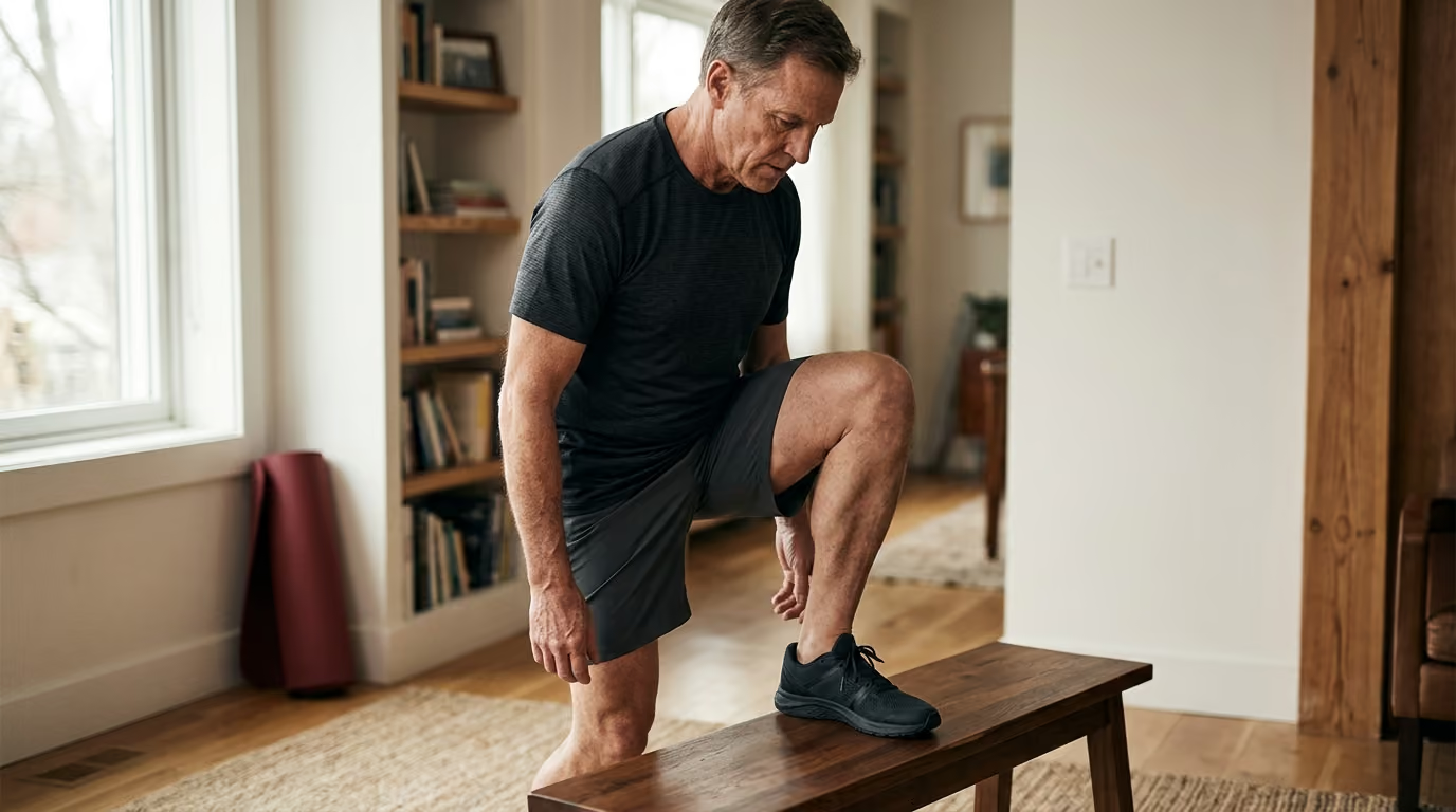
When a man over 45 struggles to sink into a full squat or reach overhead without arching his lower back, the default response is almost always to stretch. Most men assume that a stiff joint means a shortened muscle that needs pulling. This assumption is usually wrong.
Movement is not a single physical trait. When you cannot complete a movement cleanly, your body is responding to an interaction between joint structure, muscular force, movement velocity, motor coordination, pain sensitivity, and simple neurological unfamiliarity. Pulling on a muscle that your nervous system is deliberately guarding will not solve your movement problems.
Targeted assessment lets you stop guessing. By learning how to test why a movement stalls, you can apply the exact stimulus needed to restore function instead of repeating generic stretching routines that fail to produce lasting change.
Examine What Movement Science Actually Tells Us About Midlife Restrictions
Research shows that measuring how far a joint bends does not explain how well that joint functions during daily tasks. A physical therapist or trainer can use a goniometer to record the angle of your hip or shoulder. That angle tells you only the outer boundary of your passive range. It does not tell you if the limitation comes from the joint capsule, muscle stiffness, weakness, poor motor control, pain, or protective muscle guarding.
A comprehensive 2023 systematic review examined measurement tools for older adults and found a striking gap in the literature. The researchers found no validated assessment tools specifically designed to measure overall joint function in mature populations. The review noted that published studies frequently confuse passive range of motion with genuine joint function.
Being able to pull your knee toward your chest while lying on a table does not mean your hip can support your body weight during a deep lunge. Functional capability requires strength, speed, balance, and tissue tolerance working together.
Longitudinal evidence confirms that separate physical attributes drive physical independence after midlife. In a study tracking 391 community-dwelling older adults over two years, researchers evaluated leg strength, movement velocity, rapid coordination, joint range of motion, and trunk endurance as independent variables. They found that weaker leg strength increased the odds of persistent functional impairment by more than three times. Reduced trunk endurance and slower movement speed also independently predicted physical decline.
Restricted knee motion predicted functional decline as well, but its statistical impact was smaller than the effects of weakness and slow movement speed. This research demonstrates that while joint range matters, force production and movement speed are often more influential for maintaining physical capability.
Flexibility training alone rarely translates into better real-world performance. A systematic review examining healthy older adults found that while stretching routines can increase measured joint angles, the evidence that stretching improves daily functional performance is mixed and conflicting. If an awkward gait or stiff squat stems from weakness, lack of balance, or protective guarding, pulling on the muscle changes the test angle without fixing the movement.
To build a reliable movement assessment, you must stop treating mobility as one single attribute. You need a system that separates structural limitations from control problems, strength deficits, and sensory barriers.
Understand Why Movement Changes After 45 Without Blaming Normal Aging
Physical changes occur across tissues as the decades pass, but normal aging is not a disease process. Understanding the biological mechanisms behind movement changes allows you to address them directly rather than accepting stiffness as inevitable.
Connective tissues undergo structural changes over time. Collagen fibers within tendons, ligaments, and joint capsules develop more cross-links. This cross-linking increases tissue stiffness and reduces passive compliance. At the same time, tendons and fascial sheaths experience a minor decrease in water content, which alters how smoothly tissue layers glide past one another during complex movements.
These structural shifts mean that passive tissue extensibility naturally declines if tissues are not regularly loaded through full ranges of motion. However, tissue stiffness also provides joint stability and helps transfer muscular force. Increased stiffness only becomes a movement limitation when it blocks the range required for your chosen activities.
Neuromuscular changes also alter movement quality after 45. The nervous system naturally reduces the discharge rate of motor units and selectively downregulates fast-twitch muscle fibers when high-velocity demands are absent. When you stop moving quickly or lifting challenging loads, your nervous system loses its ability to recruit muscular force rapidly.
This loss of motor unit recruitment often presents as movement stiffness. If your brain senses that your muscles cannot stabilize a joint at the end of its range, it creates active muscular tension to stop you from entering that position. You feel tight, but the tightness is an active protective strategy created by your nervous system, not a short muscle.
Sensory processing and pain sensitivity change with age and activity history. Past injuries, minor joint irritation, and sedentary desk work can make the nervous system overly protective. When the brain perceives a specific movement pattern as risky, it downregulates movement amplitude long before mechanical tissue limits are reached.
Recognizing these physical and neural mechanisms changes how you approach self-assessment. A limitation in your squat or reach is rarely a permanent structural defect. It is often a combination of tissue adaptation, reduced motor recruitment, and protective neural tone that you can systematically test and improve through our mobility, joints, and functional movement resources.
Translate Assessment Principles Into Real World Performance
To make movement testing useful, you must connect clinical measurement principles to daily physical capabilities. A number on a flexibility test is meaningless if it does not help you walk, lift, climb, or play sports without limitation.
In real life, movement limitations fall into six primary categories:
- True joint capsule or mechanical restrictions where the joint surfaces cannot physically move further.
- Muscular extensibility limits where the muscle-tendon unit resists elongation under tension.
- Force production deficits where the muscles lack the strength to move the skeleton through the pattern.
- Movement velocity deficits where the body cannot generate force quickly enough to complete dynamic actions.
- Coordination and motor control gaps where the brain cannot organize the timing of multiple muscle groups.
- Protective guarding caused by pain sensitivity, fear of injury, or complete lack of recent exposure to the task.
Consider a common movement challenge: stepping onto a high ledge or climbing steep stairs while hiking. If you struggle with this movement, generic advice tells you to stretch your hip flexors. An evidence-led assessment looks at the system differently.
If you can passively pull your knee to your chest while lying on your back, your hip joint and muscles have sufficient range for the high step. If you cannot lift that same knee toward your chest while standing on one leg, your limitation is hip flexor strength or single-leg balance. If you can lift the leg easily but your knee caves inward and shakes as you push down to step up, your limitation is eccentric knee control and gluteal force production.
The same distinction applies to getting off the floor. If you find getting up from the ground awkward, you might assume your hips are too stiff. Testing may reveal that your hips move freely when supported, but you lack the trunk endurance and single-leg pushing strength required to stand up smoothly.
Applying flexibility training to a force deficit leaves you frustrated. Applying heavy resistance training to a true joint capsule restriction can lead to joint irritation. Translating assessment principles into real capability means identifying which of the six limiters is stopping you, then applying the right solution.
Separate Structural Joint Range From Muscle Extensibility
The first major diagnostic step is determining whether a movement boundary is caused by structural restrictions or an active control problem. A reliable clinical principle is to compare active movement against assisted or passive movement.
Active range of motion represents the distance your joint travels when your own muscles produce the force. Passive or assisted range of motion represents the distance your joint travels when an external force, such as a strap, a trainer, or gravity, assists the movement. Comparing these two numbers reveals how your nervous system manages joint space.
- ACTIVE VS. ASSISTED RANGE OF MOTION
- Pattern A
- Active Range: Low Assisted Range: High
- Causes: Weakness, poor coordination, protective guarding
- Pattern B
- Active Range: Low Assisted Range: Low
- Causes: Joint capsule restriction, shortened tissues
Pattern A: Active Range Is Limited, But Assisted Range Is Greater
If you can achieve a deep position with assistance but cannot reach that position under your own power, you do not have a fixed structural joint restriction. Your joint surfaces and passive connective tissues permit the movement.
This pattern indicates that your limitation comes from:
- Insufficient muscular strength in the prime movers at end range.
- Poor coordination and timing between opposing muscle groups.
- Protective neurological guarding driven by lack of stability or past pain.
- Lack of confidence and unfamiliarity with the end-range position.
When Pattern A is present, passive stretching is unhelpful. You already possess the tissue length. You need dedicated strength and muscle training and active motor control drills to teach your nervous system how to use the range you already have.
Pattern B: Active and Assisted Ranges Are Both Restricted
If you cannot achieve the desired position actively, and you still cannot reach it when an external force assists you, the restriction involves physical tissue capacity or deep protective tension.
This pattern suggests:
- Mechanical restrictions within the joint capsule or surrounding ligaments.
- Long-term structural adaptations in the muscle-tendon unit.
- High-level protective muscle guarding that does not release under passive load.
When Pattern B occurs, the next step is to examine the endpoint sensation. If the joint stops abruptly with a hard, unyielding sensation, the limit is likely bony or capsular. If the joint stops with a firm, elastic resistance, the limitation involves muscle-tendon stiffness.
Comparing both sides of your body provides critical context. If your right hip rotates 45 degrees outward while your left rotates only 15 degrees with a hard block, you may be dealing with asymmetric joint anatomy or past injury. If both sides share the same symmetrical firm boundary, you are observing your baseline structural architecture.
Pattern C: Joint Restrictions Are Consistent Across Tasks
A true structural or tissue restriction appears consistently across different exercises and body positions.
If your ankle dorsiflexion is mechanically blocked, your knee will fail to travel forward over your toes during a standing squat, a split squat, a lunge, and a seated ankle test. The restriction remains identical regardless of task complexity.
If your ankle moves forward easily during a seated test but locks up during a heavy barbell squat, your limitation is not ankle dorsiflexion. Your limitation is balance, spinal loading capacity, or squat coordination. Testing joints in simplified, low-load positions isolates structural boundaries from complex movement demands.
Practical Testing Sequence for Range Differentiation
To test any restricted movement, use this systematic approach:
- Define the exact movement error. Avoid vague statements like "my shoulders feel tight." Note the exact position: "My right arm stops ten degrees short of vertical when reaching overhead."
- Test active range without load. Stand upright and raise your arm overhead as far as you can without arching your spine. Record the stopping point.
- Test assisted range in a supported position. Lie flat on your back on the floor. Use your other hand or a light strap to gently guide the arm overhead. If your arm easily reaches the floor when lying down, you have full passive range. Your standing limitation is active stability and trunk control, not shoulder stiffness.
- Check for side-to-side asymmetries. Repeat the exact test on the opposite side. Note whether the boundary feels identical or significantly restricted on one side.
- Re-test after low-intensity movement. Perform five to ten slow, supported repetitions through the comfortable range. If the available range increases immediately after a brief warm-up, the limitation is driven by protective neural tone rather than fixed tissue shortening.
Distinguish True Muscle Weakness From Speed and Coordination Gaps
Once you determine that a movement is not blocked by a structural restriction, you must evaluate force capacity, movement velocity, and motor organization. These three physical qualities are often lumped together, but they require different assessment strategies.
In the longitudinal study of older adults cited earlier, researchers showed that leg strength, leg velocity, and coordination represented separate physical impairments. Weaker leg strength carried an odds ratio of 3.45 for persistent functional limitation. Reduced trunk extensor endurance had an odds ratio of 2.98. Slower movement velocity carried an odds ratio of 2.35. Each factor contributed independently to movement failure.
- FORCE, SPEED, AND COORDINATION TRIAGE
- Test 1: Force Capacity
- NO - Primary limiter is STRENGTH
- YES - Proceed to Test 2
- Test 2: Speed / Power
- NO - Primary limiter is POWER/SPEED
- YES - Proceed to Test 3
- Test 3: Coordination
- NO - Limiter is MOTOR CONTROL
- YES - Capacity is verified
Assessing Absolute Force Capacity
Force capacity is the ability of your muscular system to produce tension against resistance without time pressure. If you lack the base strength to move your body through a pattern slowly, you will never perform the movement well under dynamic conditions.
To assess force capacity independently of speed or balance:
- Select a supported version of the movement, such as a box squat with hand support on a rail or a slow, supported split squat.
- Perform the movement with a deliberate three-second lowering phase and a three-second rising phase.
- Observe whether your muscles can sustain smooth, continuous tension without shaking, collapsing, or requiring momentum.
If you cannot lower yourself under control or stand up without pulling with your arms, your primary limitation is muscular strength. Stretching will not improve this pattern. You need progressive resistance to build foundational force.
Assessing Movement Velocity and Power
Power is the rate at which you produce force. Many men over 45 maintain reasonable slow-speed strength from years of traditional lifting or manual work, but they lose the ability to generate force quickly.
When you lose movement speed, tasks like catching your balance after a trip, climbing stairs briskly, or changing direction become clumsy. A systematic review of physical measurement tools noted that while slow-speed strength tests are common, muscle power assessments such as the 30-second sit-to-stand power test provide unique insight into functional capacity.
To test movement velocity:
- Perform a standard movement, such as rising from a chair or completing a bodyweight heel raise, at a slow, controlled pace.
- Perform the same movement at a normal, self-selected speed.
- Attempt to perform the rising phase of the movement as rapidly and explosively as possible while maintaining balance.
If your slow-speed movement is stable and smooth, but your performance degrades dramatically when asked to move quickly, your limiting factor is rate of force development. Your training must incorporate controlled, dynamic speed work rather than just slow grinds.
Assessing Motor Coordination and Balance
Coordination is the ability of your central nervous system to synchronize multiple joints and muscle groups efficiently. Coordination deficits are frequently misdiagnosed as weakness or tightness.
A coordination or balance problem typically shows distinct characteristics:
- Inconsistent movement pathways where every repetition looks slightly different.
- Excessive upper body leaning or arm flailing to compensate for lower body instability.
- Hesitation and jerky transitions when shifting weight from one leg to the other.
- Immediate improvement in movement quality when a light fingertip touch is provided for balance.
Consider an illustrative assessment model of a man attempting a single-leg step-down from a low platform. If his knee wobbles wildly and his hip drops sideways on the first two repetitions, but stabilizes completely when he lightly touches a wall with one finger, his muscles possess adequate strength. The wall provides no mechanical lift. It provides sensory feedback that his nervous system uses to coordinate the movement.
When coordination is the limiter, loading the movement heavily or stretching the surrounding muscles will not correct the problem. The correct approach is deliberate, high-frequency practice with light support, gradually removing the sensory aids as the motor pattern solidifies.
Assess Pain Sensitivity, Fear of Movement, and Task Unfamiliarity
The brain regulates movement output based on its perception of threat. If your nervous system believes that entering a specific position will cause tissue damage, it will actively restrict your range of motion and reduce muscular output.
Research on kinesiophobia, defined as an irrational or excessive fear of physical movement resulting from vulnerability to painful injury, demonstrates how deeply psychological factors alter physical capability. A study of older adults published in physical function research revealed that greater fear of movement predicted lower daily physical activity, higher sedentary time, and worse functional performance, even in individuals without chronic pain conditions.
In older adults suffering from low back pain, research shows that kinesiophobia directly predicts impaired mobility and balance, even when lower-limb strength and grip strength remain intact. Fear changes how you use your physical capacity without necessarily reducing your raw muscular hardware.
Furthermore, a systematic review in the British Journal of Sports Medicine found strong evidence linking higher kinesiophobia to increased pain intensity and greater disability across musculoskeletal conditions. When assessing a movement limit, you must separate three distinct psychological and sensory questions:
- THE THREE-PART SENSORY AND THREAT AUDIT
- Question 1: Direct Pain
- Question 2: Anticipated Pain
- Question 3: Task Exposure
Differentiating Direct Pain From Pain Sensitivity
Pain is a complex protective output, not a direct measurement of tissue damage. If a movement causes sharp, escalating pain, your body is signaling that current load exceeds tissue tolerance. This requires medical evaluation or load reduction.
Pain sensitivity, however, behaves differently:
- Discomfort appears before any mechanical tissue boundary is reached.
- The sensation changes substantially based on your expectation or attention.
- The movement becomes completely pain-free when load is reduced by ten percent or when hand support is provided.
- Symptoms decrease after several calm, controlled repetitions.
If pain sensitivity is the primary limiter, aggressively forcing the joint into painful ranges reinforces protective guarding. Instead, you must train within a pain-free or low-discomfort window to demonstrate to your nervous system that the movement is safe.
Identifying Fear of Movement and Guarding
Fear-related limitations occur when you avoid positions because you expect them to cause harm, even if the tissues are completely healed. This is common in men over 45 who have experienced past back spasms or knee sprains.
You can identify fear-driven limitations by observing:
- Protective breath-holding and full-body bracing during light, non-threatening movements.
- Reluctance to bend the spine, flex the knees, or rotate the hips even when unweighted.
- Rapid expansion of range of motion when the movement is broken down into simple, supported steps.
Researchers frequently use validated tools like the Tampa Scale for Kinesiophobia to measure fear avoidance in clinical studies. For self-assessment, you can simply rate your confidence from zero to ten before attempting a movement. If your confidence is low despite normal muscle strength, fear and protective guarding are restricting your movement.
Addressing Lack of Task Exposure
Sometimes a movement feels awkward and stiff simply because you have not performed it in twenty years. The human body adapts specifically to the positions it occupies regularly. If your daily life involves sitting in chairs, walking on flat pavement, and lifting objects in front of your chest, your nervous system downregulates the motor pathways for deep squatting, overhead reaching, and single-leg balancing.
Research examining flexibility in mature adults aged 55 to 86 found that general physical activity levels, including light and moderate daily tasks, did not correlate with hip or shoulder flexibility. Walking ten thousand steps a day maintains cardiovascular health, but it does not preserve the specific joint angles required for deep lunges or overhead movement.
Lack of exposure is easy to identify. The movement feels unfamiliar and clumsy on the first attempt, but improves noticeably across three to four practice sets without any stretching or formal warm-up. Your body simply needs regular, graded exposure to re-learn the pattern.
Execute an Eight-Step Movement Assessment Battery
To apply these principles systematically, use this practical eight-step assessment battery. This sequence walks you through any restricted movement pattern to isolate the true limiting factor.
- THE 8-STEP MOVEMENT ASSESSMENT FLOW
- Step 1: Define the real-world target task.
- Step 2: Test an unloaded baseline version.
- Step 3: Compare active range against assisted range.
- Step 4: Test slow force capacity with external support.
- Step 5: Test movement speed and power output.
- Step 6: Test learning rate, consistency, and repeatability.
- Step 7: Audit symptoms, anticipated pain, and confidence.
- Step 8: Match the identified limiter to the correct training tool.
Step 1: Define the Real-World Target Task
Select the specific functional movement that is currently restricted. Avoid testing abstract positions that have no bearing on your physical goals. Common target tasks include:
- Lowering into a full squat with your heels on the ground.
- Reaching both arms vertically overhead without overarching your lumbar spine.
- Stepping down from an eight-inch step with control.
- Rotating your upper body to look behind you while backing up a car or swinging a club.
- Getting down to the floor and standing back up without using furniture for support.
Step 2: Test an Unloaded Baseline Version
Perform the movement at a self-selected, comfortable pace without external weights. Observe where the movement stalls, where your balance falters, or where compensatory movements occur.
Record the exact location of the limitation. For example: "During the bodyweight squat, my torso pitches forward and my heels lift when my thighs reach parallel to the floor."
Step 3: Compare Active Range Against Assisted Range
Isolate the primary joints involved in the movement and test them in supported positions.
For a squat, lie on your back and pull your knees toward your chest. If your hips flex past ninety degrees and your knees touch your ribs easily while lying down, your hips possess the physical range needed for a deep squat. If your knees stop well short of your chest with a hard block, you have a structural or tissue range restriction.
Step 4: Test Slow Force Capacity
Take balance and speed out of the equation by using external support. Hold onto a sturdy rail, wall, or suspension strap. Lower yourself slowly into the movement pattern over three seconds, hold the bottom position for two seconds, and push back up over three seconds.
If you cannot complete this slow, supported repetition smoothly, you have a force deficit. Your muscles lack the strength to stabilize and lift your body weight through that range.
Step 5: Test Speed and Power
If you pass the slow force test easily, test the movement under velocity demands. Perform the concentric, rising phase of the movement as quickly as possible while maintaining perfect control.
If your movement becomes unstable, jerky, or significantly reduced in amplitude when speed is introduced, your limitation is movement velocity and rate of force development.
Step 6: Test Learning Rate and Repeatability
Perform five continuous repetitions of the movement with clear focus and simple verbal cues. Observe whether the movement improves from the first repetition to the fifth repetition.
If your movement quality improves rapidly across repetitions, your limitation is motor coordination and lack of exposure. Your nervous system simply needs practice to organize the motor pattern.
Step 7: Audit Symptoms, Anticipated Pain, and Confidence
Rate your experience during the movement across three simple metrics:
- Pain level: Rate any physical discomfort from zero to ten during and after the movement.
- Anticipated threat: Rate your pre-movement fear or expectation of injury from zero to ten.
- Movement confidence: Rate your certainty in completing the movement safely from zero to ten.
If pain is low, but anticipated threat is high and confidence is low, protective neural guarding is actively restricting your movement.
Step 8: Match the Identified Limiter to the Correct Intervention
Once you have identified the primary constraint, select the training intervention that directly addresses it:
- For True Joint Range Limitations (Pattern B): Use targeted end-range joint mobilizations, loaded active stretching, and consult a physical therapist if sharp pain or severe asymmetry is present. Explore structured mobility and recovery strategies to support tissue health.
- For Muscular Extensibility Limits: Use eccentric strength training through the full available range rather than passive static stretching.
- For Force Production Deficits: Implement progressive resistance training using supported or scaled variations to build foundational strength.
- For Speed and Power Deficits: Introduce low-load, high-velocity movements, such as medicine ball throws, kettlebell swings, or rapid bodyweight repetitions.
- For Coordination and Balance Deficits: Use high-frequency, low-fatigue movement practice with gradual removal of external balance support.
- For Pain Sensitivity and Fear: Apply graded exposure principles, starting with non-threatening variations and slowly expanding range as confidence returns.
Avoid Common Misinterpretations and Recognize Where Evidence Is Limited
Navigating movement advice after midlife requires critical thinking. Popular fitness culture frequently misinterprets mobility research, leading men into ineffective or counterproductive routines.
Misinterpretation 1: Stretching Always Improves Functional Capability
The belief that passive flexibility automatically translates into better physical performance is not supported by research. As documented in the systematic review on flexibility in older adults, stretching interventions reliably increase joint angles on a measurement table, but their effect on real-world functional tasks is inconsistent and conflicting.
Flexibility is purely passive. Function is dynamic and active. Lengthening a hamstring through static stretching does nothing to improve your ability to hinge your hips while deadlifting or stepping over an obstacle. Range of motion must be paired with muscular control and stability to become functional.
Misinterpretation 2: General Daily Activity Maintains Joint Range
Many active men assume that walking, running, or cycling is enough to preserve joint mobility. Research contradicts this assumption. The study examining physical activity in adults aged 55 to 86 demonstrated that overall physical activity volume had no meaningful association with hip or shoulder flexibility.
Cardiovascular exercise moves your joints through narrow, repetitive mid-range arcs. Cycling never requires full hip extension. Walking never requires full overhead shoulder flexion. To maintain joint range, you must deliberately expose your joints to end-range positions through structured movement.
Misinterpretation 3: Every Movement Limit Represents Structural Damage
When a joint feels stiff or restricted, men often assume they have developed severe arthritis, cartilage tears, or permanent structural damage. While osteoarthritis and degenerative joint changes do occur with age, imaging studies consistently show that structural changes on an X-ray or MRI correlate poorly with pain and movement function.
Many individuals with significant joint changes move freely and without pain. Conversely, individuals with clean scans often experience severe stiffness due to protective neural guarding, weakness, and fear avoidance. Never assume that a movement restriction is a permanent structural sentence until you have systematically tested strength, coordination, and exposure.
Where Movement Research Remains Limited
While the principles in this guide are grounded in published evidence, it is essential to acknowledge the boundaries of current scientific literature.
First, there is no single, universally validated assessment algorithm specifically proven to diagnose all movement limitations in adults over 45. The 2023 systematic review on older-adult measurement tools highlighted that the fitness and physical therapy fields lack standardized, validated batteries for overall joint function. The assessment sequence presented here is an evidence-led clinical reasoning framework, not an automated diagnostic formula.
Second, research on muscle power and movement velocity in mature populations is less extensive than research on basic strength and balance. While tools like the 30-second sit-to-stand power test show promise, many power assessment methods are supported by single studies with small sample sizes.
Third, the measurement of kinesiophobia in older adults is complex. The 2022 review on fear avoidance in mature populations noted significant variation in how questionnaires are applied and interpreted. Questionnaires provide helpful screening data, but they cannot replace careful observation of movement behavior under real-world conditions.
Understanding these research limitations keeps your training grounded. You do not need a perfect clinical laboratory to make progress. You need a structured, observant process that helps you select the right training tools for your body, supporting your journey toward longevity science and physical optimization. For more information about our research standards, you can read about our editorial approach at Everfitguys.
Apply The Core Takeaways to Your Daily Training
Assessing movement limitations after 45 requires moving past generic stretching routines. By systematically testing whether your limitations stem from structural barriers, strength deficits, speed loss, coordination gaps, or protective guarding, you can direct your energy toward interventions that actually work.
Key Takeaways
- Differentiate Range from Function: Passive joint flexibility does not equal active functional capability.
- Compare Active and Assisted Motion: A large gap between active and passive range points to a strength, coordination, or confidence deficit rather than a structural joint restriction.
- Test Force and Speed Independently: Weakness and slow movement velocity are stronger predictors of functional decline after midlife than joint stiffness alone.
- Audit Psychological and Sensory Factors: Fear of movement, pain sensitivity, and lack of recent task exposure create active muscular tension that mimics structural stiffness.
- Match the Tool to the Limiter: Use resistance training for force deficits, speed work for velocity loss, motor practice for coordination gaps, and graded exposure for protective guarding.
Stop pulling on muscles that are trying to protect you, diagnose the true constraint in your movement system, and build the strength, control, and confidence needed to stay capable for decades to come.
Sources
- Kinesiophobia Predicts Physical Function and ... - PMC - NIH
- Kinesiophobia, Pain, Muscle Functions, and ... - PMC - NIH
- Association of kinesiophobia with pain, disability and ...
- Kinesiophobia and Fear Avoidance in Older Adults
- Flexibility Training and Functional Ability in Older Adults: A Systematic Review
- Role of kinesiophobia on pain, disability and quality of life in people suffering from chronic musculoskeletal pain: a systematic review
- Flexibility of Older Adults Aged 55–86 Years and the Influence ...
Stay sharp
Get the word
Join our newsletter for the best writing advice and stories from the field.